
Add to Cart
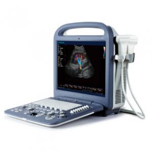
Portable Ultrasound Scannner Color Doppler Ultrasound 15 Inch LCD monitor 4D
Model:MC-DU-S2
Feasures
Ergonomic Design
Portable design, be ready at anytime and anywhere
• 15-inch high resolution LCD monitor with wide-view angle
• Standard PC keyboard, easy input
• Two probe sockets with probe holder, better protection for probes
• Rechargeable lithium battery, 1 hour scanning without power
supply
• Abundant peripherals: DICOM3.0, VGA, video out, USB, S-Video,
Footswitch etc.
Comprehensive Functions
Complete working modes, outstanding 2D performance, sensitive blood
flow imaging, 4D image technology, endows S2 with the best package
of functions in its class!
Complete working modes:
• B Mode, Dual B, 4B
• M Mode, Steer M,
• Color Mode, DPI Mode
• PW Mode, CW Mode (optional)
Advanced Imaging Technology
• THI
• μ-Scan Speckle Reduction (optional)
• Compound Imaging (optional)
• Panoramic Imaging (optional)
• Trapezoid Imaging (optional)
• 4D imaging (optional)
Broad range of clinical application
• General Practice: Abdominal, OB, GYN, Vascular, Urology, Small
Parts
• Post-measurement and calculations software packages
• Professional report for different exams
Multiple Transducers
Convex, Linear, Transvaginal, Transrectal, Intraoperative, Phased
array, Micro-convex, Volume 4D
Optimum Workflow
Upgraded workflow, smart data management, a more user-friendly and
productive Color Doppler system!
• Intuitive user interface: Clear system layout designed for
convenient workflow, Menu-driven interface
• Define your own work-style: Fully customized work settings for
data table/graph, function keys, icons, formulas, measurement,
report layout…totally based on your own work style and habit!
• Clip-board function: Easy image review during scanning
• Smart patient data management: Easy access to patient image and
data storage, retrieve, review and report.
• Value-added DICOM functions: Save, Storage Commitment, MPPS,
Print, Worklist; providing more advanced file management solutions;
• Various data solutions: built-in 320G hard drive, DICOM 3.0 LAN
port, VGA port, support Flash disk, External hard drive, external
DVD drive, video printer, USB laser/jet printer and so on.
• M-tuning: Intelligent one key image optimization
Application
♦ Liver
The ultrasonic examination of liver is a relatively high
informative method. The doctor evaluates the dimensions of the
liver, its structure and homogeneity, local disturbances as well as
the blood flow condition. The ultrasonic examination allows to
detect both diffusive changes in the liver (liver steatosis,
chronic active liver disease, cirrhosis) and local (fluid
formalities and tumor mass).
♦ The gall bladder and bile passages
Besides the liver itself, the state of gall bladder and bile
passages is also examined: their dimensions, the thickness of the
paries, patency, the presence of concrements, the state of the
connective tissue.
In most cases the ultrasonic examination allows to detect the
concrements in the gall.
♦ Pancreatic gland
During the examination of the pancreatic gland the doctors can
evaluate its dimensions, form, boundaries, homogeneity of
parenchyma, the presence of formalities.
♦ Kidney and atrabiliary capsules, retroperitoneal space
During the examination of the kidney the doctors evaluate their
number, arrangement, dimensions, form, boundaries, structure of
parenchyma and pelvicalyceal system. The ultrasonic examination
allows to detect the anomalies of the kidney, the presence of
concrements, fluid formalities and tumor mass as well as changes
resulting from inveterate and pathological lancinating processes in
kidney. The ultrasonic examination can be used for detection of the
indication of intestinal obstruction and indirect indication of
adhesive process. With the help of ultrasonic examination the
doctor can detect the presence of free liquid in the abdominal
cavity (if it is in a great number) that can play an important role
in the treatment policy of a number of therapeutic and surgical
disorders and injuries.
♦ Thyroid gland
The ultrasonic examination is the crucial in the examination of the
thyroid gland because it allows to detect the presence of
ganglions, hydatids, the changes in the dimensions and structure.
♦ Cardiology, cardiosurgery
Echocardiography is the ultrasonic diagnostics of the heart
diseases. This examination evaluates the dimensions of the heart
and its particular structures (ventricle of the heart, auricle,
ventricular septum,
thickness of ventricular myocardium, auricle etc.) the presence and
the volume of fluid in the pericard – "pericardial sac", the state
of the valves. With the help of special calculations and sizing
echocardiology allows to detect the heart mass, contractility of
the heart - ejection fraction, etc. There are special guides that
help to observe during the operation the state of the mitral valve
situated between the ventricle and auricle.
♦ Tocology, gynecology, prenatal diagnostics
The ultrasonic examination is used for examination of the internal
female genital neoplasms, the state of the gravid uterus, anatomy
and monitoring of the Embryo-fetal development. This effect is
widely used in the tocology because the sound from the venter can
be easily registered. At early stages of pregnancy the sound gets
through the bladder. When the venter is filled with the fluid it
starts to conduct the sound. The position of the placenta is
detected by the sounds of blood getting through it, and within 9-10
weeks from the moment of the fetation the doctor can listen to the
beating of its heart. The number of foetuses and the death of the
foetus can be detected with the help of ultrasound examination
♦ Muscle and skeletal examinations
The ultrasonic examination is used to detect traumatic injuries and
inflammatory disease of joints (shoulder, knee, etc.), muscles,
bands, meniscus, chorda, arthrosis.
Image